COVID Ages the Brain 10 Years
Have you had COVID? Always feel fatigued?
Have you had COVID?
Always feel fatigued?
Do you think you avoided having post-COVID issues? Maybe, maybe not … If you have any ongoing health and wellness issues, you may have Long COVID and not even know it. (source: Time 2022) New scientific data now shows 1 in 5 Americans who have had COVID still have Long COVID or other post-COVID issues.
(source: National Center for Health Statistics 2022)
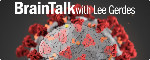
Science has studied hundreds of thousands of people infected with COVID and many others who continue to suffer from its ongoing persisting symptoms, often called Long COVID. Research shows COVID directly attacks the brain, leaving long-lasting neurological and physical issues such as brain fog and fatigue.
Your brain may have just aged 7 to 20 years.
For anyone infected by COVID, even if they are not seriously ill, the infection is equivalent to aging the brain by 7 to 10 years. For those that experience COVID with brain fog, it can be equivalent to up to 20 years! Remember, your brain begins shrinking in your 30s or 40s and dramatically increases the shrinkage rate by the time you are in your 60s. COVID highly accelerates this.
Initial symptoms are just the tip of the iceberg. Below the surface, there may be cascading issues that range from … loss of cognitive abilities like brain fog, memory loss, and poor focus/concentration … to psychological problems like anxiety and depression … to physical problems like loss of taste & smell, chronic pain (chest, muscle, joints, headaches), shortness of breath or difficulty breathing, persistent cough, fast or pounding heartbeat, fever, dizziness, insomnia, fatigue and much more. Other health issues may become worse after physical or mental activity.
See what Cereset can do for post Covid brain fog.
Hear what actual Cereset® clients have to say about how our process has helped their memory, cognitive, and metal clarity issues resulting from Covid 19.
Have you lost hope?
Cereset can help.
Cereset is likely to help the brain help itself to gain regulation and lessen or eliminate these long-COVID symptoms since the brain drives the body. A harmonized brain can help itself to likely reverse these harmful and frightening issues caused by COVID. How do you know if your brain is out of harmony? How do you get it back into harmony? Cereset of course … find a center near you!


Cereset can help your brain help you resolve COVID issues.
Most Long COVID symptoms are likely the results of a brain traumatized by the virus. Cereset has developed a new protocol from working with brain data of previously infected COVID clients. Our new protocol can now support the brain to restore itself and lessen the brain’s accelerated aging effects. Cereset has been proven to help relax your brain, so it resets itself to likely mitigate these Long COVID symptoms. Cereset has helped over a hundred thousand others with their health and wellness challenges, as well as been studied in many clinical trials. This is an exciting time for Cereset, and all of the Cereset offices across the country are available to assist you.
Cereset’s patented BrainEcho® technology is non-invasive, safe, proven, and highly effective in helping the brain relax to support brain-driven anti-aging. Cereset helps the brain reset itself to its natural state, restoring rhythm and harmony. Clients typically experience a reduction of symptoms in as little as four to five sessions in less than three weeks.
Science Confirms COVID Can Attack You Any Time After Your COVID Infection

Your brain may be under attack!
Attack leaves long-lasting neurological issues … source: A Sobering Addition to the Literature on COVID-19 and the Brain (Yale University via JCI). Other research found virus levels were over 1,000 times higher in the brain than in the body … source: Neuroinvasion and Encephalitis Following Inoculation of SARS-CoV-2 in K18-hACE2 Mice (Georgia State University via Science Immunology). Cereset has been proven to help reverse this process and protect the brain.

Aging Happens with COVID Infections!
COVID infections age the brain by 7 to 10 years. COVID infections which include the symptom of brain fog, age the brain by 20 years … source: Cognitive Impairment from Severe COVID-19 Equivalent to 20 Years of Aging (University of Cambridge via Neuroscience news). Cereset has an advanced brain-driven anti-aging protocol that can help your brain overcome this accelerated brain aging process.

Experienced Brain Fog? It may be the COVID you just had.
COVID causes cognitive impairment, often experienced as “brain fog”, similar to what goes on with the brain over 50 and 70 years of age and “is the equivalent to losing 10 IQ points” … source: Cognitive Impairment From Severe COVID-19 Equivalent to 20 Years of Aging (University of Cambridge). If you have had any type of cognitive impairment (brain fog or dementia) from COVID, Cereset can help restore your lost IQ AND enable you to recover to a brain capability beyond where you were when COVID began.

Long COVID is caused by the brain.
Long COVID issues experienced in the body are actually driven by the brain … source: How Effects on the Brain Can Produce Long COVID (Yale School of Medicine). Cereset understands that the brain drives the body, and so this is true! Cereset can help as one of the only currently known solutions to the COVID brain problem.
Our Latest News

Scottsdale, Arizona
Cereset Antiaging™ – a brain-driven antiaging™ solution
The Problem
We spend over $60 Billion each year on antiaging products and services in the USA. Obviously, it is vital that we stay as young as possible (in appearance and health). Yet, the real source of aging – the brain – is not being addressed successfully. Understanding antiaging from the brain’s perspective is critical.
The brain’s cerebral cortex (outer layer) begins to shrink when we are about 30 years of age. As we grow older, the brain shrinks faster, beginning at about age 60, and for those headed toward Alzheimer’s Disease or Dementia, the brain shrinks very fast in some specific areas. (reported by the Center of Lifespan Changes of Oslo, Norway).

Scottsdale, Arizona
Cereset – The Greatest Weapon Against Long COVID and an Aging Brain
COVID has infected over 458 million people around the world. OMICRON – the newest COVID-like virus – has infected over 130 million people globally, is much more infectious, and has a much higher death rate. Science has identified an ongoing issue with all of those who COVID has infected – these infected individuals, even if they do not realize it, have a brain that has become smaller. Any COVID infection is a significant str…

Scottsdale, Arizona
Cereset Efficacy Proven In Relationship to Bio-Functions
It is amazing when we realize that half of us in the US as adults will hear the words – “you have cancer.” One out of two of us will hear these words and will never be the same for the remainder of our life. Each of us has a 50/50 chance to hear it in our lifetime – wow, that is truly significant.
Cancer is a disease that we need to be aware of so we can best understand how to stay healthy, how to resist cancer during the cancer…

Scottsdale, Arizona
Cereset Efficacy Proven In Relationship to Bio-Functions
For over a decade, Cereset and its legacy technology have been confirmed by measuring biological functions to measure efficacy. In everything that has been measured to date, Cereset is shown to have set the gold standard to support the brain’s own correction of many issues.
HRV (heart rate variability) along with BP (blood pressure) has enabled researchers to see the efficacy of Cereset for sleep, PTSD, hot flashes…
References to explore
Clinical course and outcomes of critically ill patients with SARS-CoV-2 pneumonia in Wuhan, China: A single-centered, retrospective, observational study
X. Yang et al., Clinical course and outcomes of critically ill patients with SARS-CoV-2 pneumonia in Wuhan, China: A single-centered, retrospective, observational study. Lancet Respir. Med. 8, 475–481 (2020).
Extrapulmonary manifestations of COVID-19
A. Gupta et al., Extrapulmonary manifestations of COVID-19. Nat. Med. 26, 1017–1032 (2020).
Post-acute COVID-19 syndrome
A. Nalbandian et al., Post-acute COVID-19 syndrome. Nat. Med. 27, 601–615 (2021).
COVID-19: ICU delirium management during SARS-CoV-2 pandemic
K. Kotfis et al., COVID-19: ICU delirium management during SARS-CoV-2 pandemic. Crit. Care 24, 176 (2020).
Neurologic manifestations of hospitalized patients with coronavirus disease 2019 in Wuhan, China
L. Mao et al., Neurologic manifestations of hospitalized patients with coronavirus disease 2019 in Wuhan, China. JAMA
oroNerve Study Group, Neurological and neuropsychiatric complications of COVID-19 in 153 patients: A UK-wide surveillance study
A. Varatharaj et al; CoroNerve Study Group, Neurological and neuropsychiatric complications of COVID-19 in 153 patients: A UK-wide surveillance study. Lancet Psychiatry 7, 875–882 (2020).
Cerebral micro-structural changes in COVID-19 patients – An MRI-based 3-month follow-up study
Y. Lu et al., Cerebral micro-structural changes in COVID-19 patients – An MRI-based 3-month follow-up study. EClinicalMedicine 25, 100484 (2020).
Clinical course and risk factors for mortality of adult inpatients with COVID-19 in Wuhan, China: A retrospective cohort study
F. Zhou et al., Clinical course and risk factors for mortality of adult inpatients with COVID-19 in Wuhan, China: A retrospective cohort study. Lancet 395, 1054–1062 (2020).
Psychosocial factors and hospitalisations for COVID-19: Prospective cohort study based on a community sample
G. D. Batty et al., Psychosocial factors and hospitalisations for COVID-19: Prospective cohort study based on a community sample. Brain Behav. Immun. 89, 569–578 (2020).
Functional and cognitive outcomes after COVID-19 delirium
B. C. Mcloughlin et al., Functional and cognitive outcomes after COVID-19 delirium. Eur. Geriatr. Med. 11, 857–862 (2020).
Neurological manifestations and COVID-19: Experiences from a tertiary care center at the Frontline
P. Pinna et al., Neurological manifestations and COVID-19: Experiences from a tertiary care center at the Frontline. J. Neurol. Sci. 415, 116969 (2020).
Severe acute respiratory syndrome coronavirus 2 (SARS-CoV-2) and the central nervous system
. G. De Felice, F. Tovar-Moll, J. Moll, D. P. Munoz, S. T. Ferreira, Severe acute respiratory syndrome coronavirus 2 (SARS-CoV-2) and the central nervous system. Trends Neurosci. 43, 355–357 (2020).
Detection of severe acute respiratory syndrome coronavirus in the brain: Potential role of the chemokine mig in pathogenesis
J. Xu et al., Detection of severe acute respiratory syndrome coronavirus in the brain: Potential role of the chemokine mig in pathogenesis. Clin. Infect. Dis. 41, 1089–1096 (2005).
Possible central nervous system infection by SARS coronavirus.
K.-K. Lau et al., Possible central nervous system infection by SARS coronavirus. Emerg. Infect. Dis. 10, 342–344 (2004).
Detection of SARS coronavirus RNA in the cerebrospinal fluid of a patient with severe acute respiratory syndrome
E. C. W. Hung et al., Detection of SARS coronavirus RNA in the cerebrospinal fluid of a patient with severe acute respiratory syndrome. Clin. Chem. 49, 2108–2109 (2003).
A first case of meningitis/encephalitis associated with SARS-Coronavirus-2
T. Moriguchi et al., A first case of meningitis/encephalitis associated with SARS-Coronavirus-2. Int. J. Infect. Dis. 94, 55–58 (2020).
SARS-CoV-2 detected in cerebrospinal fluid by PCR in a case of COVID-19 encephalitis
Y. H. Huang, D. Jiang, J. T. Huang, SARS-CoV-2 detected in cerebrospinal fluid by PCR in a case of COVID-19 encephalitis. Brain Behav. Immun. 87, 149 (2020).
Two patients with acute meningoencephalitis concomitant with SARS-CoV-2 infection
R. Bernard-Valnet et al., Two patients with acute meningoencephalitis concomitant with SARS-CoV-2 infection. Eur. J. Neurol. 27, e43–e44 (2020).
Magnetic resonance imaging alteration of the brain in a patient with coronavirus disease 2019 (COVID-19) and anosmia
L. S. Politi, E. Salsano, M. Grimaldi, Magnetic resonance imaging alteration of the brain in a patient with coronavirus disease 2019 (COVID-19) and anosmia. JAMA Neurol. 77, 1028–1029 (2020).
Neuropathology of COVID-19: A spectrum of vascular and acute disseminated encephalomyelitis (ADEM)-like pathology
R. R. Reichard et al., Neuropathology of COVID-19: A spectrum of vascular and acute disseminated encephalomyelitis (ADEM)-like pathology. Acta Neuropathol. 140, 1–6 (2020).
Neuropathology of patients with COVID-19 in Germany: A post-mortem case series
J. Matschke et al., Neuropathology of patients with COVID-19 in Germany: A post-mortem case series. Lancet Neurol. 19, 919–929 (2020).
Neuroinvasion of SARS-CoV-2 in human and mouse brain
E. Song et al., Neuroinvasion of SARS-CoV-2 in human and mouse brain. bioRxiv [Preprint] (2020).
https://www.biorxiv.org/content/10.1101/2020.06.25.169946v2 (Accessed 8 November 2020)
Lethality of SARS-CoV-2 infection in K18 human angiotensin-converting enzyme 2 transgenic mice
F. S. Oladunni et al., Lethality of SARS-CoV-2 infection in K18 human angiotensin-converting enzyme 2 transgenic mice. Nat. Commun. 11, 6122 (2020).
SARS-CoV-2 infection impacts carbon metabolism and depends on glutamine for replication in Syrian hamster astrocytes
G. de Oliveira et al., SARS-CoV-2 infection impacts carbon metabolism and depends on glutamine for replication in Syrian hamster astrocytes. bioRxiv [Preprint] (2021).
https://www.biorxiv.org/content/10.1101/2021.10.23.465567v1 (Accessed 30 October 2021)
SARS-CoV-2 targets neurons of 3D human brain organoids
A. Ramani et al., SARS-CoV-2 targets neurons of 3D human brain organoids. EMBO J. 39, e106230 (2020).
SARS-CoV-2 infects human neural progenitor cells and brain organoids
B.-Z. Zhang et al., SARS-CoV-2 infects human neural progenitor cells and brain organoids. Cell Res. 30, 928–931 (2020).
COVID-19 treatments and pathogenesis including anosmia in K18-hACE2 mice
J. Zheng et al., COVID-19 treatments and pathogenesis including anosmia in K18-hACE2 mice. Nature 589, 603–607 (2021).
SARS-CoV-2 crosses the blood-brain barrier accompanied with basement membrane disruption without tight junctions alteration
L. Zhang et al., SARS-CoV-2 crosses the blood-brain barrier accompanied with basement membrane disruption without tight junctions alteration. Signal Transduct. Target. Ther. 6, 337 (2021).
Long Covid: Una nuova sfida oltre l’emergenza
A. Grieco, Long Covid: Una nuova sfida oltre l’emergenza. Come ritrovarebenessere e salute dopo il Covid-19 (Naturvis Books, 2021).
SARS-CoV-2 infects the brain choroid plexus and disrupts the blood-CSF barrier in human brain organoids
L. Pellegrini et al., SARS-CoV-2 infects the brain choroid plexus and disrupts the blood-CSF barrier in human brain organoids. Cell Stem Cell 27, 951–961.e5 (2020).
The SARS-CoV-2 spike protein alters barrier function in 2D static and 3D microfluidic in-vitro models of the human blood-brain barrier
T. P. Buzhdygan et al., The SARS-CoV-2 spike protein alters barrier function in 2D static and 3D microfluidic in-vitro models of the human blood-brain barrier. Neurobiol. Dis. 146, 105131 (2020).
SARS-COV2 alters blood brain barrier integrity contributing to neuro-inflammation
J. L. Reynolds, S. D. Mahajan, SARS-COV2 alters blood brain barrier integrity contributing to neuro-inflammation. J. Neuroimmune Pharmacol. 16, 4–6 (2021).
Prevalence and impact of cardiovascular metabolic diseases on COVID-19 in China
B. Li et al., Prevalence and impact of cardiovascular metabolic diseases on COVID-19 in China. Clin. Res. Cardiol. 109, 531–538 (2020).
Double-stranded RNA is produced by positive-strand RNA viruses and DNA viruses but not in detectable amounts by negative-strand RNA viruses
F. Weber, V. Wagner, S. B. Rasmussen, R. Hartmann, S. R. Paludan, Double-stranded RNA is produced by positive-strand RNA viruses and DNA viruses but not in detectable amounts by negative-strand RNA viruses. J. Virol. 80, 5059–5064 (2006).
Establishment of a Brazilian line of human embryonic stem cells in defined medium: Implications for cell therapy in an ethnically diverse population
A. M. Fraga et al., Establishment of a Brazilian line of human embryonic stem cells in defined medium: Implications for cell therapy in an ethnically diverse population. Cell Transplant. 20, 431–440 (2011).
Neutralizing antibody and soluble ACE2 inhibition of a replication-competent VSV-SARS-CoV-2 and a clinical isolate of SARS-CoV-2
J. B. Case et al., Neutralizing antibody and soluble ACE2 inhibition of a replication-competent VSV-SARS-CoV-2 and a clinical isolate of SARS-CoV-2. Cell Host Microbe 28, 475–485.e5 (2020).
Dysregulation of brain and choroid plexus cell types in severe COVID-19
A. C. Yang et al., Dysregulation of brain and choroid plexus cell types in severe COVID-19. Nature 595, 565–571 (2021).
Neuropilin-1 is a host factor for SARS-CoV-2 infection
J. L. Daly et al., Neuropilin-1 is a host factor for SARS-CoV-2 infection. Science 370, 861–865 (2020).
Neuropilin-1 facilitates SARS-CoV-2 cell entry and infectivity
L. Cantuti-Castelvetri et al., Neuropilin-1 facilitates SARS-CoV-2 cell entry and infectivity. Science 370, 856–860 (2020).
CD147-spike protein is a novel route for SARS-CoV-2 infection to host cells. Signal Transduct
K. Wang et al., CD147-spike protein is a novel route for SARS-CoV-2 infection to host cells. Signal Transduct. Target Ther. 5, 283 (2020).
No evidence for basigin/CD147 as a direct SARS-CoV-2 spike binding receptor
J. Shilts, T. W. M. Crozier, E. J. D. Greenwood, P. J. Lehner, G. J. Wright, No evidence for basigin/CD147 as a direct SARS-CoV-2 spike binding receptor. Sci. Rep. 11, 413 (2021).
Astroglial metabolic networks sustain hippocampal synaptic transmission
N. Rouach, A. Koulakoff, V. Abudara, K. Willecke, C. Giaume, Astroglial metabolic networks sustain hippocampal synaptic transmission. Science 322, 1551–1555 (2008).
Neurotoxic reactive astrocytes are induced by activated microglia
S. A. Liddelow et al., Neurotoxic reactive astrocytes are induced by activated microglia. Nature 541, 481–487 (2017).
Block of A1 astrocyte conversion by microglia is neuroprotective in models of Parkinson’s disease
S. P. Yun et al., Block of A1 astrocyte conversion by microglia is neuroprotective in models of Parkinson’s disease. Nat. Med. 24, 931–938 (2018).
Knockout of reactive astrocyte activating factors slows disease progression in an ALS mouse model
K. A. Guttenplan et al., Knockout of reactive astrocyte activating factors slows disease progression in an ALS mouse model. Nat. Commun. 11, 3753 (2020).
Supply and demand in cerebral energy metabolism: The role of nutrient transporters
I. A. Simpson, A. Carruthers, S. J. Vannucci, Supply and demand in cerebral energy metabolism: The role of nutrient transporters. J. Cereb. Blood Flow Metab. 27, 1766–1791 (2007).
Astrocytes are poised for lactate trafficking and release from activated brain and for supply of glucose to neurons
G. K. Gandhi, N. F. Cruz, K. K. Ball, G. A. Dienel, Astrocytes are poised for lactate trafficking and release from activated brain and for supply of glucose to neurons. J. Neurochem. 111, 522–536 (2009).
SARS-CoV-2 is associated with changes in brain structure in UK Biobank
G. Douaud et al., SARS-CoV-2 is associated with changes in brain structure in UK Biobank. Nature 604, 697–707 (2022).
Long-term microstructure and cerebral blood flow changes in patients recovered from COVID-19 without neurological manifestations
Y. Qin et al., Long-term microstructure and cerebral blood flow changes in patients recovered from COVID-19 without neurological manifestations. J. Clin. Invest. 131, e147329 (2021).
The role of the orbitofrontal cortex in anxiety disorders
M. R. Milad, S. L. Rauch, The role of the orbitofrontal cortex in anxiety disorders. Ann. N. Y. Acad. Sci. 1121, 546–561 (2007).
Plasma biomarkers of neuropathogenesis in hospitalized patients with COVID-19 and those with postacute sequelae of SARS-CoV-2 infection
B. A. Hanson et al., Plasma biomarkers of neuropathogenesis in hospitalized patients with COVID-19 and those with postacute sequelae of SARS-CoV-2 infection. Neurol. Neuroimmunol. Neuroinflamm. 9, 9 (2022).
Are we facing a crashing wave of neuropsychiatric sequelae of COVID-19? Neuropsychiatric symptoms and potential immunologic mechanisms
E. A. Troyer, J. N. Kohn, S. Hong, Are we facing a crashing wave of neuropsychiatric sequelae of COVID-19? Neuropsychiatric symptoms and potential immunologic mechanisms. Brain Behav. Immun. 87, 34–39 (2020).
Psychiatric and neuropsychiatric presentations associated with severe coronavirus infections: A systematic review and meta-analysis with comparison to the COVID-19 pandemic
J. P. Rogers et al., Psychiatric and neuropsychiatric presentations associated with severe coronavirus infections: A systematic review and meta-analysis with comparison to the COVID-19 pandemic. Lancet Psychiatry 7, 611–627 (2020).
COVID-19 induces neuroinflammation and loss of hippocampal neurogenesis
R. Klein et al., COVID-19 induces neuroinflammation and loss of hippocampal neurogenesis. Research Square [Preprint] (2021). https://www.researchsquare.com/article/rs-1031824/v1 (Accessed 5 November 2021).
A human three-dimensional neural-perivascular ‘assembloid’ promotes astrocytic development and enables modeling of SARS-CoV-2 neuropathology
L. Wang et al., A human three-dimensional neural-perivascular ‘assembloid’ promotes astrocytic development and enables modeling of SARS-CoV-2 neuropathology. Nat. Med. 27, 1600–1606 (2021).
Dysregulation of brain and choroid plexus cell types in severe COVID-19
A. C. Yang et al., Dysregulation of brain and choroid plexus cell types in severe COVID-19. Nature 595, 565–571 (2021).
SARS-CoV-2 is not detected in the cerebrospinal fluid of encephalopathic COVID-19 patients
D. G. Placantonakis et al., SARS-CoV-2 is not detected in the cerebrospinal fluid of encephalopathic COVID-19 patients. Front. Neurol. 11, 587384 (2020).
Human pluripotent stem cell-derived neural cells and brain organoids reveal SARS-CoV-2 neurotropism predominates in choroid plexus epithelium
F. Jacob et al., Human pluripotent stem cell-derived neural cells and brain organoids reveal SARS-CoV-2 neurotropism predominates in choroid plexus epithelium. Cell Stem Cell 27, 937–950 (2020).
Update on neurological manifestations of COVID-19
H. Yavarpour-Bali, M. Ghasemi-Kasman, Update on neurological manifestations of COVID-19. Life Sci. 257, 118063 (2020).
Tropism of SARS-CoV-2 for developing human cortical astrocytes
M. G. Andrews et al., Tropism of SARS-CoV-2 for developing human cortical astrocytes. bioRxiv [Preprint] (2021). https://www.biorxiv.org/content/10.1101/2021.01.17.427024v1 (Accessed 18 March 2021).
Olfactory transmucosal SARS-CoV-2 invasion as a port of central nervous system entry in individuals with COVID-19.
J. Meinhardt et al., Olfactory transmucosal SARS-CoV-2 invasion as a port of central nervous system entry in individuals with COVID-19. Nat. Neurosci. 24, 168–175 (2021).
Neurological complications associated with the blood-brain barrier damage induced by the inflammatory response during SARS-CoV-2 infection
I. Alquisiras-Burgos et al., Neurological complications associated with the blood-brain barrier damage induced by the inflammatory response during SARS-CoV-2 infection. Mol. Neurobiol. 58, 520–535 (2021).
SARS-CoV-2 cell entry depends on ACE2 and TMPRSS2 and is blocked by a clinically proven protease inhibitor
M. Hoffmann et al., SARS-CoV-2 cell entry depends on ACE2 and TMPRSS2 and is blocked by a clinically proven protease inhibitor. Cell 181, 271–280.e8 (2020).
ApoE-isoform-dependent SARS-CoV-2 neurotropism and cellular response
C. Wang et al., ApoE-isoform-dependent SARS-CoV-2 neurotropism and cellular response. Cell Stem Cell 28, 331–342.e5 (2021).
Multilevel proteomics reveals host perturbation strategies of SARS-CoV-2 and SARS-CoV
A. Stukalov et al., Multilevel proteomics reveals host perturbation strategies of SARS-CoV-2 and SARS-CoV. Nature 594, 246–252 (2021).
Proteomics of SARS-CoV-2-infected host cells reveals therapy targets
D. Bojkova et al., Proteomics of SARS-CoV-2-infected host cells reveals therapy targets. Nature 583, 469–472 (2020).
Shotgun proteomics analysis of SARS-CoV-2-infected cells and how it can optimize whole viral particle antigen production for vaccines
L. Grenga et al., Shotgun proteomics analysis of SARS-CoV-2-infected cells and how it can optimize whole viral particle antigen production for vaccines. Emerg. Microbes Infect. 9, 1712–1721 (2020).
Sweet sixteen for ANLS
L. Pellerin, P. J. Magistretti, Sweet sixteen for ANLS. J. Cereb. Blood Flow Metab. 32, 1152–1166 (2012).
Neuroprotective role of lactate after cerebral ischemia
C. Berthet et al., Neuroprotective role of lactate after cerebral ischemia. J. Cereb. Blood Flow Metab. 29, 1780–1789 (2009).
Astrocyte-neuron lactate transport is required for long-term memory formation
A. Suzuki et al., Astrocyte-neuron lactate transport is required for long-term memory formation. Cell 144, 810–823 (2011).
Knockout of GAD65 has major impact on synaptic GABA synthesized from astrocyte-derived glutamine
A. B. Walls et al., Knockout of GAD65 has major impact on synaptic GABA synthesized from astrocyte-derived glutamine. J. Cereb. Blood Flow Metab. 31, 494–503 (2011).
Role of glutamate in neuron-glia metabolic coupling
P. J. Magistretti, Role of glutamate in neuron-glia metabolic coupling. Am. J. Clin. Nutr. 90, 875S–880S (2009).
The role of astroglia in neuroprotection
M. Bélanger, P. J. Magistretti, The role of astroglia in neuroprotection. Dialogues Clin. Neurosci. 11, 281–295 (2009).
Brain energy metabolism: Focus on astrocyte-neuron metabolic cooperation
M. Bélanger, I. Allaman, P. J. Magistretti, Brain energy metabolism: Focus on astrocyte-neuron metabolic cooperation. Cell Metab. 14, 724–738 (2011).
Sugar for the brain: The role of glucose in physiological and pathological brain function
P. Mergenthaler, U. Lindauer, G. A. Dienel, A. Meisel, Sugar for the brain: The role of glucose in physiological and pathological brain function. Trends Neurosci. 36, 587–597 (2013).
The glutamate/GABA-glutamine cycle: Aspects of transport, neurotransmitter homeostasis and ammonia transfer
L. K. Bak, A. Schousboe, H. S. Waagepetersen, The glutamate/GABA-glutamine cycle: Aspects of transport, neurotransmitter homeostasis and ammonia transfer. J. Neurochem. 98, 641–653 (2006).
Glutamate released spontaneously from astrocytes sets the threshold for synaptic plasticity
C. Bonansco et al., Glutamate released spontaneously from astrocytes sets the threshold for synaptic plasticity. Eur. J. Neurosci. 33, 1483–1492 (2011).
18F-FDG brain PET hypometabolism in patients with long COVID
E. Guedj et al., 18F-FDG brain PET hypometabolism in patients with long COVID. Eur. J. Nucl. Med. Mol. Imaging 48, 2823–2833 (2021).
FDG PET signal is driven by astroglial glutamate transport
E. R. Zimmer et al., [18F]FDG PET signal is driven by astroglial glutamate transport. Nat. Neurosci. 20, 393–395 (2017).
Astrocyte biomarkers in Alzheimer’s disease
S. F. Carter et al., Astrocyte biomarkers in Alzheimer’s disease. Trends Mol. Med. 25, 77–95 (2019).
Glucose utilization: Still in the synapse
A. J. Stoessl, Glucose utilization: Still in the synapse. Nat. Neurosci. 20, 382–384 (2017).
Long-term neuropsychiatric complications and 18F-FDG-PET hypometabolism in the brain from prolonged infection of COVID-19
A. T. Yu, N. M. Absar, Long-term neuropsychiatric complications and 18F-FDG-PET hypometabolism in the brain from prolonged infection of COVID-19. Alzheimer Dis. Assoc. Disord. 36, 173–175 (2021).
Long COVID: Cognitive complaints (brain fog) and dysfunction of the cingulate cortex
J. Hugon, E.-F. Msika, M. Queneau, K. Farid, C. Paquet, Long COVID: Cognitive complaints (brain fog) and dysfunction of the cingulate cortex. J. Neurol. 269, 44–46 (2022).
About the source and consequences of 18F-FDG brain PET hypometabolism in short and long COVID-19
I. C. Fontana, D. G. Souza, L. Pellerin, D. O. Souza, E. R. Zimmer, About the source and consequences of 18F-FDG brain PET hypometabolism in short and long COVID-19. Eur. J. Nucl. Med. Mol. Imaging 48, 2674–2675 (2021).
Cognitive impairment and altered cerebral glucose metabolism in the subacute stage of COVID-19
J. A. Hosp et al., Cognitive impairment and altered cerebral glucose metabolism in the subacute stage of COVID-19. Brain 144, 1263–1276 (2021).
Neurotoxic reactive astrocytes drive neuronal death after retinal injury
K. A. Guttenplan et al., Neurotoxic reactive astrocytes drive neuronal death after retinal injury. Cell Rep. 31, 107776 (2020).
Neurotoxic reactive astrocytes induce cell death via saturated lipids
K. A. Guttenplan et al., Neurotoxic reactive astrocytes induce cell death via saturated lipids. Nature 599, 102–107 (2021).
Cortical microstructural correlates of astrocytosis in autosomal-dominant Alzheimer disease
E. Vilaplana et al., Cortical microstructural correlates of astrocytosis in autosomal-dominant Alzheimer disease. Neurology 94, e2026–e2036 (2020).
Decreased number and increased activation state of astrocytes in gray and white matter of the prefrontal cortex in autism
G. Vakilzadeh et al., Decreased number and increased activation state of astrocytes in gray and white matter of the prefrontal cortex in autism. Cereb. Cortex, https://doi.org/10.1093/cercor/bhab523 (2022).
Cell type-specific manifestations of cortical thickness heterogeneity in schizophrenia
M. A. Di Biase et al., Cell type-specific manifestations of cortical thickness heterogeneity in schizophrenia. Mol. Psychiatry 27, 2052–2060. (2022).
Cortical thickness differences are associated with cellular component morphogenesis of astrocytes and excitatory neurons in nonsuicidal self-injuring youth
S. Cai et al., Cortical thickness differences are associated with cellular component morphogenesis of astrocytes and excitatory neurons in nonsuicidal self-injuring youth. Cereb. Cortex, https://doi.org/10.1093/cercor/bhac103 (2022).
The neuroanatomy of autism spectrum disorder: An overview of structural neuroimaging findings and their translatability to the clinical setting
C. Ecker, The neuroanatomy of autism spectrum disorder: An overview of structural neuroimaging findings and their translatability to the clinical setting. Autism 21, 18–28 (2017).
Development of cortical thickness and surface area in autism spectrum disorder
V. T. Mensen et al., Development of cortical thickness and surface area in autism spectrum disorder. Neuroimage Clin. 13, 215–222 (2016).
“Epilepsy pathology” in Encyclopedia of the Neurological Sciences
M. Thom, “Epilepsy pathology” in Encyclopedia of the Neurological Sciences, R. B. Daroff, M. J. Aminoff, Eds. (Elsevier, 2014), pp. 136–141.
Reactive astrocyte nomenclature, definitions, and future directions
C. Escartin et al., Reactive astrocyte nomenclature, definitions, and future directions. Nat. Neurosci. 24, 312–325 (2021).
Reduced expression of glutamate transporters vGluT1, EAAT2 and EAAT4 in learned helpless rats, an animal model of depression
M. Zink, B. Vollmayr, P. J. Gebicke-Haerter, F. A. Henn, Reduced expression of glutamate transporters vGluT1, EAAT2 and EAAT4 in learned helpless rats, an animal model of depression. Neuropharmacology 58, 465–473 (2010).
Astroglial correlates of neuropsychiatric disease: From astrocytopathy to astrogliosis.
R. Kim, K. L. Healey, M. T. Sepulveda-Orengo, K. J. Reissner, Astroglial correlates of neuropsychiatric disease: From astrocytopathy to astrogliosis. Prog. Neuropsychopharmacol. Biol. Psychiatry 87 (Pt A), 126–146 (2018).
Astrogliosis and episodic memory in late life: Higher GFAP is related to worse memory and white matter microstructure in healthy aging and Alzheimer’s disease.
B. M. Bettcher et al., Astrogliosis and episodic memory in late life: Higher GFAP is related to worse memory and white matter microstructure in healthy aging and Alzheimer’s disease. Neurobiol. Aging 103, 68–77 (2021).
Data from “Morphological, cellular, and molecular basis of drain infection in COVID-19 patients.”
F. Crunfli et al., Data from “Morphological, cellular, and molecular basis of drain infection in COVID-19 patients.” PRIDE partner repository. Deposited 25 January 2021.
https://www.ebi.ac.uk/pride/archive?keyword=PXD023781
As you learn more about the importance of a harmonized brain, be sure to schedule an Introduction to Cereset special with your nearest client center.
This 50-minute evaluation includes an orientation with your personal Tech Coach, a baseline brain observation to assess your brain’s harmony with ability to manage stress, your very own Personal Brain Index®, and a recommended plan of action. Please check with your local client center for special pricing on this offer.


